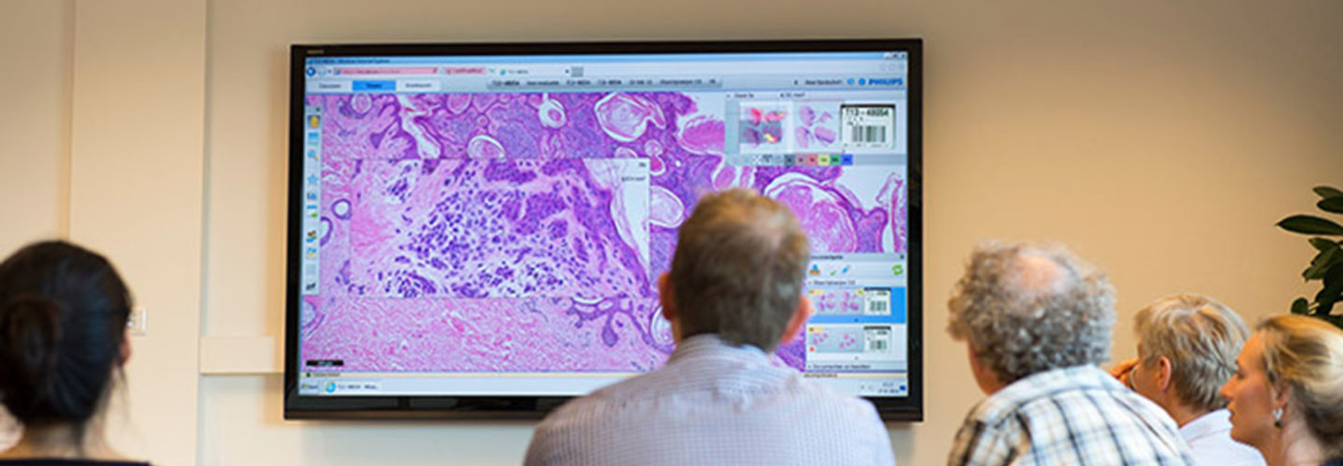
Deep Learning Networks Help Pathologists Zoom In on Cancer Biopsies
Deep-learning computer networks are giving breast cancer diagnoses a boost.
A research team from Case Western Reserve University’s Center of Computational Imaging and Personalized Diagnostics recently published a study that detailed a deep-learning network approach to diagnose breast cancer. The department built, trained and analyzed a computer network to determine whether a machine could more accurately evaluate biopsies.
The research team first trained the network using 400 samples of tissue from different sites, and then presented the network with 200 cases from The Cancer Genome Atlas.
According to the results of the study, published in Scientific Reports, the network was able to accurately determine the presence of cancer on whole slides with 100 percent accuracy.
“The experimental results show that the method is able to detect invasive breast cancer regions on whole-slide histopathology images with a high degree of precision, even when tested on cases from a cohort different to the one used for training,” the study found.
Furthermore, the method can be applied to whole-slide images to detect regions of invasive tissue; these images can be integrated with other computerized solutions in digital pathology, like tumor grading, according to the study.
“Pathologists are extremely busy, and we’re talking about microscopic-level detail in these tissue slides. So, clearly, for them to go in and pick out every cell of cancer was not tenable. There just wasn’t enough time for them to be able to sit down and manually do that,” Anant Madabhushi, professor of biomedical engineering at Case Western Reserve University and co-author of the study, tells Information Management.
The Machine Making Digital Pathology Possible
The network can be more sophisticated, granular and accurate than pathologists in human-machine comparisons because it is nearly impossible for pathologists to determine “microscopic-level detail” in the tissue slides, Madabhushi says.
But a new device promises to make digital pathology available to pathologists. The Food and Drug Administration recently approved the marketing of Philips IntelliSite Pathology Solution, a whole-slide imaging system that allows pathologists to review and interpret digital surgical pathology slides prepared from biopsied tissue at resolutions equal to 400 times magnification.
"A pathologist can look at an image of a slide on their computer monitor, and that is equivalent to the pathologist looking at a slide under their microscope,” Madabhushi tells Information Management of the new solution. “That means digital pathology — the digitization of slides — can now be used for primary diagnosis by a pathologist. That’s a game changer.”
This new system allows pathologists to make a digital diagnosis, rather than look directly at tissues under a conventional microscope — a major step in the quest to more accurately and easily diagnose cancer, said Alberto Gutierrez, director of the Office of In Vitro Diagnostics and Radiological Health in the FDA’s Center for Devices and Radiological Health, in a statement around the new solution.
“Because the system digitizes slides that would otherwise be stored in physical files, it also provides a streamlined slide storage and retrieval system that may ultimately help make critical health information available to pathologists, other health care professionals and patients faster,” Gutierrez adds.


