
HEALTHTECH: How are cadavers used at the ARC different from those used in traditional learning environments?
Barral: We use plastinated cadavers. They’re not plastic, but actual human cadavers that are heavily embalmed and custom-made for our school. Unlike lightly embalmed cadavers that last weeks, plastinated specimens can last for years. The beauty of this approach is we can teach anatomy piecemeal. Students learn the fundamentals at the beginning. They become acquainted with them not only in the first year of their studies, but throughout the entirety of their medical education. We have full-body, predissected cadavers and multiple specimens of organs, limbs and regions.
LEARN MORE: Find out how VR simulation training is set to change nursing education.
HEALTHTECH: How does it help students learn?
Barral: The actual skills that you utilize to dissect cadavers are not in any way translated into surgical skills. Surgical skills aren't taught during dissection. You spend a lot of time, literally hours upon hours, dissecting skin and fat. And because they are lightly embalmed, you have to do it in a short amount of time. A traditional center that uses real bodies and dissects and destroys them as they go along has to procure bodies every year. They leak, they have to be in tanks, they have to be in solutions that are toxic to some people. I’ve had students wear respirators while they are dissecting because they are sensitive to chemicals.
HEALTHTECH: How has this resource been used during the pandemic?
Barral: The plans for the center, and procurement and collection, happened before the pandemic, and it didn’t lead us to modify our plans. I was hired three years ago, in July 2018. We started designing the building and did a tour of several universities and technologies to find out what was right for us. We ordered the collection of plastinated specimens, which has since grown to hundreds. The building opened just in time for the first cohort, and we started class in July 2020.
We do have the capacity to livestream, with two cameras designed for virtual teaching. We have the perfect lighting and can zoom in with great detail into any kind of structure. Everyone has been able to benefit from the center. Due to physical distancing restrictions during the pandemic, we couldn’t always get the whole class in here. So, in addition to livestreaming, we established office hours so students could come in and learn on their own when they wanted.
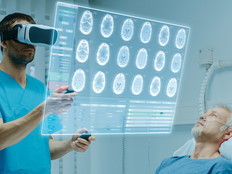
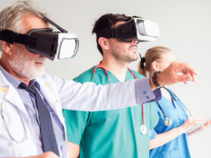
HEALTHTECH: Walk us through the technology at the center. What role do augmented and virtual reality and 3D scanners play?
Barral: We can do dissections virtually, and it takes one second to remove the skin. It’s a case where the students wouldn’t have learned anything in an hour or two by doing that. The 3D scan we use is also pretty unique. Each of the scanners has a QR code associated with it, so the students come in, use their phones, scan the specimen and get what they need. They can pull this image when they’re at the clinic, look at an angiogram after surgery, and explore it in any way with the specimen. Every structure here is labeled. It allows the for self-directed learning, which we encourage. Students need to do more of that as they become physicians, so the technology is perfectly suited for it.
READ MORE: Learn how surgeons use VR technology to train and adapt.
HEALTHTECH: How do these technologies help students understand the human body and prepare for the future?
Barral: The real educational value is that the students can learn anatomy piecemeal —longitudinally, as we say — and be exposed to it repeatedly. This spaced learning, pedologically speaking, has been demonstrated to improve understanding. And you can only do that with a permanent collection like this.
HEALTHTECH: Do you believe this is the future for medical education?
Barral: Yes. This approach has resistance from some, more traditional anatomists. I get it. I love to dissect cadavers and think it’s very interesting. But the students often forget what they’ve learned and spend more time dissecting. And there's no chance to go back because the cadaver is destroyed and no longer there. What we have to teach in medical school increases every few years, the curriculum gets larger. But we don’t get more years of teaching, so the teaching has to become more efficient.